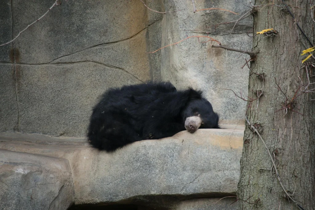
Hibernation biology reveals something far stranger than prolonged sleep. When a Japanese black bear settles into its winter den, its heart rate plummets 38% while body temperature drops only 4%[s]. This sequence matters: heart rate drops first, body temperature follows[s]. The animal is not passively cooling down. It is actively orchestrating a controlled physiological shutdown, and researchers have identified one brainstem circuit that helps make this possible.
What Hibernation Biology Actually Looks Like
Forget the image of a groggy bear taking a long nap. Hibernation biology involves a coordinated collapse of normal metabolic function that would kill most mammals. Arctic ground squirrels drop their body temperature to negative 2°C[s] and survive eight to nine months without eating or drinking[s]. In thirteen-lined ground squirrels, blood glucose plummets from 8.5 mM to 3.3 mM as cells switch from burning carbohydrates to burning fat[s].
Bears operate differently. Japanese black bears maintain body temperatures around 35.2°C during hibernation while their hearts slow to roughly 45 beats per minute[s]. Prolonged immobility and slow blood flow would raise clotting risk in non-hibernating mammals. Hibernating bears instead shift toward reversible anti-thrombotic physiology: circulating platelets and clotting factors fall during torpor, helping prevent clots until normal hemostasis returns after arousal[s].
The Brain’s Hibernation Switch
Researchers have located a specific cluster of neurons that appears to control torpor entry. Catecholaminergic neurons in the ventrolateral medulla, a region of the brainstem, become active before animals enter hibernation[s]. When scientists artificially activated these neurons in non-fasting mice, the animals entered a torpor-like state with reduced body temperature, heart rate, and energy expenditure. When they inhibited these neurons, 30% of fasting mice failed to enter torpor at all.
These VLM-CA neurons are conserved across species. A comparable VLM-CA neuron population also appears in Daurian ground squirrels[s], suggesting hibernation biology relies on an ancient neural architecture that multiple mammal lineages have preserved or reactivated.
The Muscle Paradox
Here is what makes hibernation biology medically relevant: hibernating animals emerge with their muscles intact. Humans begin losing muscle mass within a week of inactivity, and ICU patients can lose more than 10% of their muscle in seven days[s]. Yet bears wake up fit after five months of immobility.
When researchers examined gene expression in hibernating bears, they expected muscle-building genes to be suppressed to conserve energy. The opposite occurred: scores of genes involved in muscle protein biosynthesis were upregulated in a coordinated, metabolically costly pattern[s].
Where do hibernating bears get the nutrients to build muscle while fasting? Evidence points to nitrogen recycling: gut microbes may convert urea into amino acids, feeding muscle synthesis without food intake[s].
Medical Applications
Hibernation biology has direct applications to human medicine. Bears reversibly toggle insulin resistance with no health consequences[s], a feat that could inform diabetes treatment if replicated. Hibernating animals tolerate ischemia-reperfusion injury, the damage that occurs when blood flow returns to oxygen-deprived tissues after stroke or cardiac arrest[s].
Cold-induced proteins like RBM3 protect neurons during the temperature drops of torpor[s]. After hibernation, bullfrogs show improved neural function under hypoxia across the entire central nervous system[s]. Together, they point to broad neuroprotective adaptations.
Space agencies have supported research into hibernation-like states. A bearlike state of hibernation could reduce cargo requirements for Mars missions: slowed metabolism means less food, less oxygen, less fuel[s].
Climate Threats
The same physiology that makes hibernation biology remarkable also makes hibernators vulnerable to climate change. Warming den temperatures shorten torpor bouts, increase arousal frequency, and deplete the fat reserves animals need to survive winter and reproduce successfully[s].
The phenological timing of hibernation, emergence, and reproduction is becoming decoupled from environmental cues[s]. Many hibernating species already operate near their physiological limits in extreme environments, leaving little margin for adaptation as climate variability intensifies.
Physiological Mechanics of Hibernation Biology
Hibernation biology encompasses distinct physiological strategies across taxa. Large hibernators like bears undergo mild hypothermia: Japanese black bears maintain mean body temperatures of 35.2°C (95% CI 35.00-35.38) while heart rate stabilizes at 44.76 bpm (95% CI 34.54-54.98)[s]. The temporal sequence is significant: heart rate decline precedes body temperature reduction[s], indicating active cardiovascular suppression rather than passive thermodynamic cooling.
Small hibernators achieve far more extreme states. Thirteen-lined ground squirrels drop body temperature to 5°C; arctic ground squirrels reach negative 2°C[s]. Arctic ground squirrels can hibernate eight to nine months without food or water intake[s], surviving through complete metabolic reorganization.
Neural Circuitry of Torpor Induction
The neural mechanisms governing torpor have recently come into focus. Catecholaminergic neurons in the ventrolateral medulla (VLM-CA neurons) are required for normal fasting-induced torpor and sufficient to induce a torpor-like state in mice[s]. Chemogenetic inhibition of VLM-CA neurons prevented 30% of fasted mice from entering torpor and delayed torpor onset in those that did enter. Chemogenetic activation in non-fasted mice induced a torpor-like state with reduced body temperature, heart rate, O2 consumption, CO2 production, and calculated energy expenditure.
VLM-CA neurons project to the dorsal motor vagal nucleus (regulating heart rate) and medial preoptic area (regulating thermogenesis), providing the anatomical substrate for coordinated cardiovascular and thermoregulatory suppression. These neurons are conserved in Daurian ground squirrels and become active before hibernation[s], suggesting the circuit is phylogenetically ancient.
Molecular Mediators of Cold Adaptation
Hibernation biology involves major metabolic rewiring. Energy utilization shifts from carbohydrates to fatty acids: serum glucose in thirteen-lined ground squirrels drops from 8.5 mM (summer active) to 3.3 mM (torpor), while d-β-hydroxybutyrate rises from 0.26 mM to 2.3 mM[s].
Cold-shock RNA-binding proteins play critical neuroprotective roles. RBM3 transcript levels increase in liver, heart, and brain tissues during torpor in golden-mantled squirrels and black bears[s]. Cold-inducible RNA-binding protein (CIRBP) is upregulated in skeletal muscle, liver, and brown adipose tissue of thirteen-lined ground squirrels during torpor. These proteins regulate mRNA stability and translation under hypothermic conditions.
Uncoupling proteins (UCP1, UCP2, UCP3) show tissue-specific upregulation during hibernation. UCP2 transcripts increase 1.6-fold in white adipose tissue; UCP3 increases 3-fold in skeletal muscle of arctic ground squirrels[s]. HDAC1 and HDAC4 protein levels increase in skeletal muscle and brown adipose tissue during torpor, indicating epigenetic regulation.
Muscle Preservation Through IGF Signaling
Hibernating mammals resist disuse muscle atrophy despite months of immobility[s]. Gene expression analysis revealed paradoxical upregulation of scores of muscle protein biosynthesis genes in a coordinated, metabolically costly pattern[s].
IGF1 and IGF2 transcript levels are upregulated during torpor in skeletal muscle of thirteen-lined ground squirrels. In hibernating brown bears, while circulating IGF-1 and IGF-2 plasma levels decrease, tissue availability increases due to reduced Acid Labile Subunit (ALS), maintaining the anabolic IGF/IGFBP ratio in target tissues. Nitrogen recycling via gut microbiome conversion of urea to amino acids may supply the substrate for ongoing protein synthesis[s].
Translational Applications
Hibernation biology offers direct templates for drug discovery. Hibernation provides natural examples of reversible insulin resistance, hypothermia, and metabolic suppression with applications to diabetes management and ischemia-reperfusion injury[s]. Bears toggle insulin resistance seasonally without pathological consequences[s].
Hibernators tolerate hypoxia and ischemia-reperfusion stresses that are clinically relevant to stroke and cardiac arrest. The ability to emerge from hibernation without neuronal, muscle, or bone loss highlights avenues for treating neurodegenerative and age-related diseases[s]. Post-hibernation bullfrogs show improved neural function under hypoxia across the entire CNS[s].
A bearlike hibernation state could reduce resource requirements for extended space missions: slowed metabolism means less food, oxygen, and consequently less fuel[s]. In 2025, National Geographic reported that NASA and ESA were supporting studies of hibernation-like states in humans.
Climate Change Impacts on Hibernation Phenology
Elevated hibernacula temperatures shorten torpor bouts, increase arousal frequency, and deplete energy reserves crucial for survival and reproductive success[s]. The phenological timing of hibernation, emergence, and reproduction is becoming decoupled from environmental cues, creating mismatches that threaten fitness and survival[s].
Many hibernating species already inhabit extreme environments and operate near physiological limits. As climate variability intensifies, high-latitude species face ecological disruptions including habitat loss, predation shifts, and disrupted food webs, while tropical hibernators face direct physiological stress as conditions approach their thermal maxima.