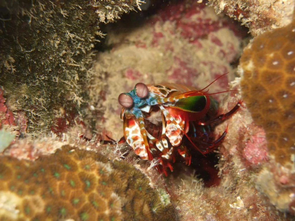
Some mantis shrimp species have 16 or more photoreceptor types. Humans normally have three color cone types. The obvious conclusion is that mantis shrimp see a visual world far richer than anything we can imagine. This conclusion is wrong, and understanding why it is wrong reveals something fundamental about how animal color vision actually works.
The Photoreceptor Myth
In the early 2010s, a popular webcomic helped spread the idea that mantis shrimp experience colors beyond human comprehension. The claim became common in popular science: more photoreceptors equals richer color experience.
Then researchers at the University of Queensland put it to the test. In a 2014 study published in Science, Hanne Thoen and colleagues trained mantis shrimp to associate specific wavelengths with food rewards, then measured whether the animals could distinguish between colors at various wavelength separations. The results contradicted the viral claim: behavioral discrimination thresholds were roughly 12 to 25nm, far coarser than human trichromatic wavelength discrimination under optimal conditions.[s][s]
This is a popular explanation that gets the science backwards. The animal with many more photoreceptor types was being outperformed at color discrimination by an animal with three.
Why More Receptors Can Mean Less
Animal color vision depends not on how many photoreceptors an eye contains, but on what the brain does with the signals those receptors send. Humans use a system called opponent-process color coding. The brain compares signals from the three cone types against each other: red versus green, blue versus yellow, light versus dark. These comparisons are continuous, detecting tiny shifts in the balance of cone activation.
The colors we see are the result of a computation, a comparison of the inputs from the different cones. Neurons literally add and subtract cone inputs to build “opponent channels.”[s] This is why three receptor types can support fine color discrimination.
Mantis shrimp do something different. Their color receptor classes appear to work less like inputs into an opponent-process comparison and more like a bank of narrow spectral filters.[s] Instead of computing fine gradations, the mantis shrimp brain appears to read which filters are active, essentially scanning a spectral barcode. This system may trade precision for speed. In the chaotic reef environment where mantis shrimp hunt, rapid recognition of prey and threats may matter more than distinguishing between slightly different shades of blue.
What Mantis Shrimp Actually See
The debunking of the color richness myth should not obscure what makes mantis shrimp vision genuinely extraordinary. They detect ultraviolet light, which the human lens normally filters out, extending their usable spectrum into wavelengths humans cannot normally see.[s]
Most remarkably, mantis shrimp can detect circular polarization, a rare visual ability best documented in stomatopods. Some species also reflect circularly polarized light, which may create a private signaling channel for other stomatopods and remain invisible to many other animals.[s]
The History Written in Our Eyes
Human color vision carries the signature of an evolutionary bottleneck. Early vertebrates possessed a broad complement of cone opsins enabling tetrachromatic or trichromatic vision. However, during the Mesozoic era, ancestral mammals underwent a nocturnal bottleneck, with a reduction in cone diversity and a predominantly rod-based, dichromatic visual system.[s]
For much of the Mesozoic, many mammalian ancestors occupied nocturnal niches. They did not need elaborate color vision to survive in dim light. Two cone types were enough. When some mammalian lineages eventually returned to daytime activity, most retained this dichromatic inheritance. Dogs, cats, and most mammals still see the world in roughly the same limited palette as their Mesozoic ancestors.
Primates took a different path. Through genetic evolution, primates developed trichromatic vision by adding green opsins responsible for medium wavelengths, thereby achieving a richer visual perception.[s] This re-evolution of color vision happened through gene duplication and spectral divergence, a case of how evolution shapes sensory systems according to ecological need. The result likely aided detection of ripe fruit against green foliage, an advantage for tree-dwelling primates.
When Losing Color Helps
Not all animal color vision has followed the path of adding receptors. Sharks demonstrate the opposite: they lost their ability to see blue and violet light entirely, and this loss was adaptive.
Ancestral cartilaginous fishes retained only the green-light-sensitive cone opsin gene and the long-wavelength-sensitive cone opsin gene, with all cartilaginous fishes losing the shortwave-sensitive opsins SWS1 and SWS2.[s] Research using zebrafish models revealed why: in the presence of SWS1 and SWS2, blue and violet light can induce cell aging, followed by photoreceptor layer thinning. The loss aids in preventing shortwave light damage to the eye.[s]
Most sharks today are likely cone monochromats, unable to see color at all.[s] In deep or turbid water where their tapetum lucidum amplifies whatever light reaches them, color information carries less survival value than maximizing light sensitivity.
The Greenland shark has taken this adaptation further. Adapted to exceptionally dim Arctic deep-sea habitats and a lifespan of up to 400 years, this species retains functional rod-based dim-light vision, but many bright-light cone-based vision genes have become pseudogenized and are no longer expressed.[s] Evolution trimmed what was not needed.
The Limits of Human Tetrachromacy
If animal color vision depends on computation as much as receptor count, could humans ever expand their color perception? The genetics suggest a narrow possibility. Tetrachromacy means that a visual system relies on four cone types. In humans, this possibility exists only in women, because the L and M opsin genes sit on the X chromosome, and women have two of them.[s]
Women who carry one normal opsin gene and one shifted variant could, in principle, express four distinct cone types. Researchers tested 24 women with this genetic setup. Only one showed clear evidence of using all four cones in color perception.[s] The brain’s visual processing system, evolved for three inputs, does not automatically exploit a fourth. Hardware alone does not determine function.
From Shrimp to Surgery
The true lesson of animal color vision is finding practical application. Researchers have engineered a camera that mimics the mantis shrimp’s ability to separate multiple wavelengths of light in compact space. By capturing ultraviolet, visible, and near-infrared light on a single chip, this bio-inspired device is designed to combine lymph-node localization with a readout of whether tissue appears suspicious during surgery.[s]
In ex vivo tests on breast cancer specimens, covering 94 lymph nodes from 33 patients, the camera’s ultraviolet readout identified cancerous lymph nodes with 97 percent sensitivity and 89 percent specificity.[s] The mantis shrimp-inspired imaging approach shows promise for rapid tissue classification, but the reported system had not yet been tested in a living organism.
Understanding how different species solve the problem of extracting information from light, rather than assuming more receptors mean better vision, leads to technologies that work with biology rather than against it. The next time you encounter a claim that some animal sees colors beyond human imagination, ask instead: what computation does that visual system perform, and what problem does it solve?
The Photoreceptor Myth
In the early 2010s, a popular webcomic described mantis shrimp (stomatopods, particularly Odontodactylus scyllarus) as experiencing colors beyond human comprehension. The claim became common in popular science: more photoreceptors equals richer color experience.
Thoen et al. (2014) put this to rigorous behavioral testing in a study published in Science. They trained mantis shrimp to associate specific wavelengths with food rewards, then measured discrimination thresholds. Behavioral wavelength discrimination thresholds fell in the roughly 12-25nm range, far coarser than human trichromatic wavelength discrimination under optimal conditions.[s][s]
This is a popular explanation that gets the science backwards. The animal with many more photoreceptor types was being outperformed at color discrimination by an animal with three.
Opponent Processing Versus Spectral Barcoding
Animal color vision depends on the neural computation applied to photoreceptor outputs, not receptor count. Human vision employs opponent-process color coding, first described by Ewald Hering in 1878 and confirmed neurophysiologically in the mid-20th century. The three cone types (S, M, L, with peak sensitivities at approximately 420nm, 530nm, and 560nm) feed into three opponent channels: L-M (red-green), S-(L+M) (blue-yellow), and L+M (luminance).
The colors we see are the result of a computation, a comparison of the inputs from the different cones. Neurons literally add and subtract cone inputs to build “opponent channels.”[s] These difference signals are continuous, enabling detection of tiny wavelength shifts.
Mantis shrimp use a fundamentally different architecture. Their color receptor classes appear to work less like inputs into an opponent-process comparison and more like a parallel bank of narrow spectral filters.[s] Each color receptor class has a narrow tuning curve and fires when its specific wavelength band is present. Rather than computing fine gradations, the stomatopod brain appears to read which filters are active. This spectral barcode system may trade resolution for recognition speed, an optimization for the dynamic, turbulent reef environment where rapid prey identification matters more than wavelength precision.
Genuine Stomatopod Visual Specializations
The debunking of the color richness myth should not obscure mantis shrimp’s legitimate visual achievements. Their ultraviolet-sensitive receptor classes extend sensitivity to roughly 300nm to 400nm, below the range normally available to human vision.[s]
Most remarkably, stomatopods can detect circular polarization, a rare visual ability best documented in this group. Some species also reflect circularly polarized light, which may create a private signaling channel for other stomatopods and remain invisible to many other animals.[s]
The Nocturnal Bottleneck and Opsin Diversity
Human color vision carries the signature of the Mesozoic nocturnal bottleneck. Early vertebrates possessed a broad complement of cone opsins enabling tetrachromatic or trichromatic vision. However, during the Mesozoic era, ancestral mammals underwent a nocturnal bottleneck, with a reduction in cone diversity and a predominantly rod-based, dichromatic visual system.[s] SWS2 (blue-sensitive) and RH2 (green-sensitive) opsins were lost, leaving only SWS1 and M/LWS.
In primates, trichromatic vision re-emerged independently through gene duplication and spectral divergence of the medium/long-wavelength-sensitive (M/LWS) opsin, marking a major evolutionary transition toward high-acuity color vision under diurnal light conditions.[s] This duplication event in catarrhine primates exemplifies how evolution shapes sensory systems. The OPN1MW and OPN1LW genes now sit adjacent on the X chromosome, sharing nearly 98% sequence identity, which explains the prevalence of red-green color blindness via unequal recombination.
Adaptive Opsin Loss in Elasmobranchs
Sharks demonstrate that evolutionary optimization can proceed by subtraction. Ancestral chondrichthyans retained only the green-light-sensitive cone opsin gene (rh2) and the long-wavelength-sensitive cone opsin gene (lws), with all cartilaginous fishes losing sws1 and sws2.[s]
Zebrafish models with SWS knockout revealed the mechanism: in the presence of SWS1 and SWS2, blue and violet light can induce cell aging, followed by photoreceptor layer thinning. The loss aids in preventing shortwave light damage to the eye.[s] This selective advantage, combined with the light-amplifying tapetum lucidum that makes color breadth less critical, may have favored SWS loss.
Sharks (Selachii) have retained only rh1 and one cone opsin gene; as such, they may be cone monochromats, lacking the ability to see colors.[s] Rays (Batoidea) retain rh2 and lws, conferring possible dichromacy.
The Greenland shark (Somniosus microcephalus) represents a deep-sea case. Genomic and transcriptomic analysis revealed that dim-light (rod-based) vision genes are intact and robustly expressed, while many bright-light (cone-based) vision genes have become pseudogenized and/or are no longer expressed.[s] The single functional visual opsin gene rh1 remains intact, while rh2, gnat2, pde6c, cnga3, and cngb3 show pseudogenization.[s] In exceptionally dim Arctic deep-sea habitats, photopic vision genes become dispensable.
Constraints on Human Tetrachromacy
If animal color vision depends on computation as much as receptor count, could humans expand their perceptual dimensionality? The genetics suggest a narrow possibility. Tetrachromacy means that a visual system relies on four cone types. In humans, this possibility exists only in women, because the L and M opsin genes sit on the X chromosome, and women have two of them.[s]
Women heterozygous for anomalous trichromacy may express S, M, normal L, and spectrally-shifted L or M, a theoretical four-channel system. Jordan et al. (2010) tested 24 women with this genotype behaviorally. Only one showed clear evidence of using all four cones in color perception.[s] The visual cortex, having evolved for three-channel input, does not automatically exploit additional channels. Neural architecture constrains perception beyond raw receptor count.
This constraint also manifests in sex differences in color discrimination. Under moderate time pressure, females outperform males by 19.889 to 29.926 points in total error scores on color discrimination tests, with the authors interpreting the result through a framework that includes photopigment variation, experience-dependent plasticity, and decision strategies.[s]
Bio-Inspired Multi-Spectral Imaging
Understanding animal color vision mechanisms enables biomimetic engineering. Gruev et al. at the University of Illinois developed a camera inspired by mantis shrimp multi-spectral architecture. The device captures ultraviolet, visible, and near-infrared light on a single chip using pixel-level filters with stacked light-sensing layers.[s]
In ex vivo testing on breast cancer specimens covering 94 lymph nodes from 33 patients, the camera’s ultraviolet readout identified cancerous nodes with 97 percent sensitivity and 89 percent specificity.[s] The spectral barcode approach, less useful for appreciating color gradients, shows promise for rapid categorical tissue classification, though the reported system had not yet been tested in a living organism.
The translational lesson: understanding what computation different visual systems perform, rather than assuming more receptors mean better vision, leads to technologies that leverage biological optimization strategies. Animal color vision research reveals not a hierarchy of richness but a diversity of solutions, each fitted to specific ecological and computational constraints.